

A 7-year-old crossbred dog, living in Hong Kong, was presented with unilateral epistaxis, with no response to medication. A CT scan revealed a small foreign body-associated lesion in the nasal cavity. At rhinoscopy, no obvious foreign body or mass was detected. A biopsy of the nasal mucosa was performed and sent for histopathology.
Histopathology of the samples of nasal mucosa revealed the process to be most likely of thromboembolic nature, resulting in extensive submucosal haemorrhage. Admixed with free blood clots were nematode larvae consistent with microfilaria.
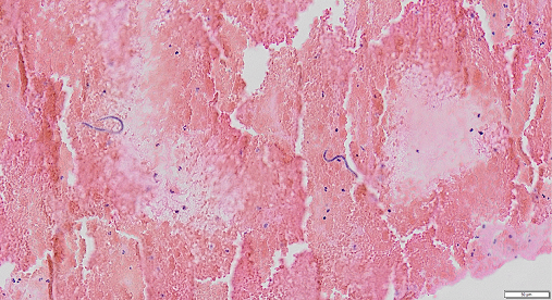
Tissue from nasal mucosa with extensive haemorrhage and two microfilarial larvae.

Higher magnification of a microfilaria larvum
QUESTIONS:
- The microfilariae are consistent with which nematode and disease?
- What diagnostic technique(s) would enable a definitive diagnosis of the disease?
Written by Dr. Ana R. Resendes
Click for the Answer
- The microfilaria detected is consistent with either Dirofilaria or Dipetalonema Dirofilaria sp. causes disease in dogs and other animal species, known as Heartworm. Dirofilaria sp. may cause zoonotic infection. Both occult and symptomatic dirofilariosis has been documented in dogs from Asia, including the Hong Kong area. In this case, although no further testing was performed, the presence of clinical signs (epixtasis), suggestive of peripheral thromboembolism, points towards an infection with Dirofilaria sp., rather than Dipetalonema reconditum, which rarely causes significant disease.
- Based on their size and shape it is possible to differentiate Dirofilaria sp. from Dipetalonema reconditum in blood smears under the microscope. However, tests based on detection of microfilaraemia do not detected occult dirofilariosis, in which there is no microfilaraemia present. Further testing for detection of antibodies and antigens may be necessary in this case (Table 1).

Discussion:
1. In dogs, various species of Dirofilaria have been reported. Dogs are the main reservoirs of immitis and D. repens. Cats may also be infected by D. repens or D. Immitis; however, cats are rarely considered to be reservoirs since microfilaraemia is rare in cats, but they can still can display clinical signs. The intermediate hosts are mosquitoes, predominantly of the genera Aedes, Culex, or Anopheles.
2. Immitis can be found in 10% of free-roaming dogs in Hong Kong. A new species of Dirofilaria has been found in both humans and dogs in Hong Kong and named “Ca. Dirofilaria hongkongensis”; however, the pathogenicity and pathology of this species in dogs or other animal hosts is unknown.
3. In dogs, adult immitis worms live in the pulmonary arteries and right chambers of the heart, but they can develop in various other arteries and veins, and have been reported from a variety of unusual sites, including the eye and brain.
4. Occult dirofilariasis, resulting from infection without microfilaraemia, occurs in 10-67% of infected dogs. Clinical diagnosis of occult dirofilariasis then rests on the findings of the indirect fluorescent antibody test, antigen detection tests, and cardiopulmonary imaging.
5. Heartworm disease is usually seen in dogs that are older than 5 years and have had continuous or multiple infections. The clinical signs are usually those of cardiovascular dysfunction, such as cough, dyspnoea, ascites and lethargy, which may progress to congestive heart failure. Right-sided heart failure develops as a consequence of pulmonary hypertension. Severe pulmonary arterial disease often results in chronic or low-grade disseminated intravascular coagulation, and hence thrombocytopenia and haemoglobinuria. Epistaxis is not frequently described, but can also be present in dogs resulting from peripheral thromboembolism in the presence of microfilaraemia. Renal failure is also a possible outcome resulting from an immune-mediated glomerulonephritis. Aberrant migration of adult heartworms into the systemic arterial circulation, a rare event, results in thromboembolism and hence signs such as hindlimb lameness and interdigital ischaemic necrosis. Renal failure is a possible outcome.
6. Vena cava syndrome (venae cavae syndrome, liver failure syndrome) is a variant of heartworm disease seen usually in young dogs in which large numbers of adult worms (100+) fill the right atrium and venae cavae. The syndrome is characterized by sudden onset of weakness, anorexia, bilirubinuria, haemoglobinuria, azotaemia and anaemia. The shock occurs because of obstruction and decreased venous return.
7. Dirofilariosis in cats usually causes cough and dyspnoea, but may also cause vomiting, neurological signs, and malaise; sudden death is another possible outcome. Vena cava syndrome also occurs in cats, and, if untreated, has a poor prognosis and high mortality rate.

Life cycle of Dirofilaria
Further Reading:
- A Novel Dirofilaria Species Causing Human and Canine Infections in Hong Kong. J Clin Microbiol. 2012 Nov; 50(11): 3534–3541.
- Guidelines for clinical management of canine Heartworm disease. European Society for Dirofilariosis and Angiostrongylosis. https://www.esda.vet/wp-content/uploads/2017/11/GUIDELINES-FOR-CLINICAL-MANAGEMENT-OF-CANINE-HEARTWORM-DISEASE.pdf
- Jubb, Kennedy & Palmer’s Pathology of Domestic Animals. 6th Edition. Cardiovascular system. Volume 3
Recent Comments